Mastitis in Cows: Signs, Causes, Treatment and Prevention
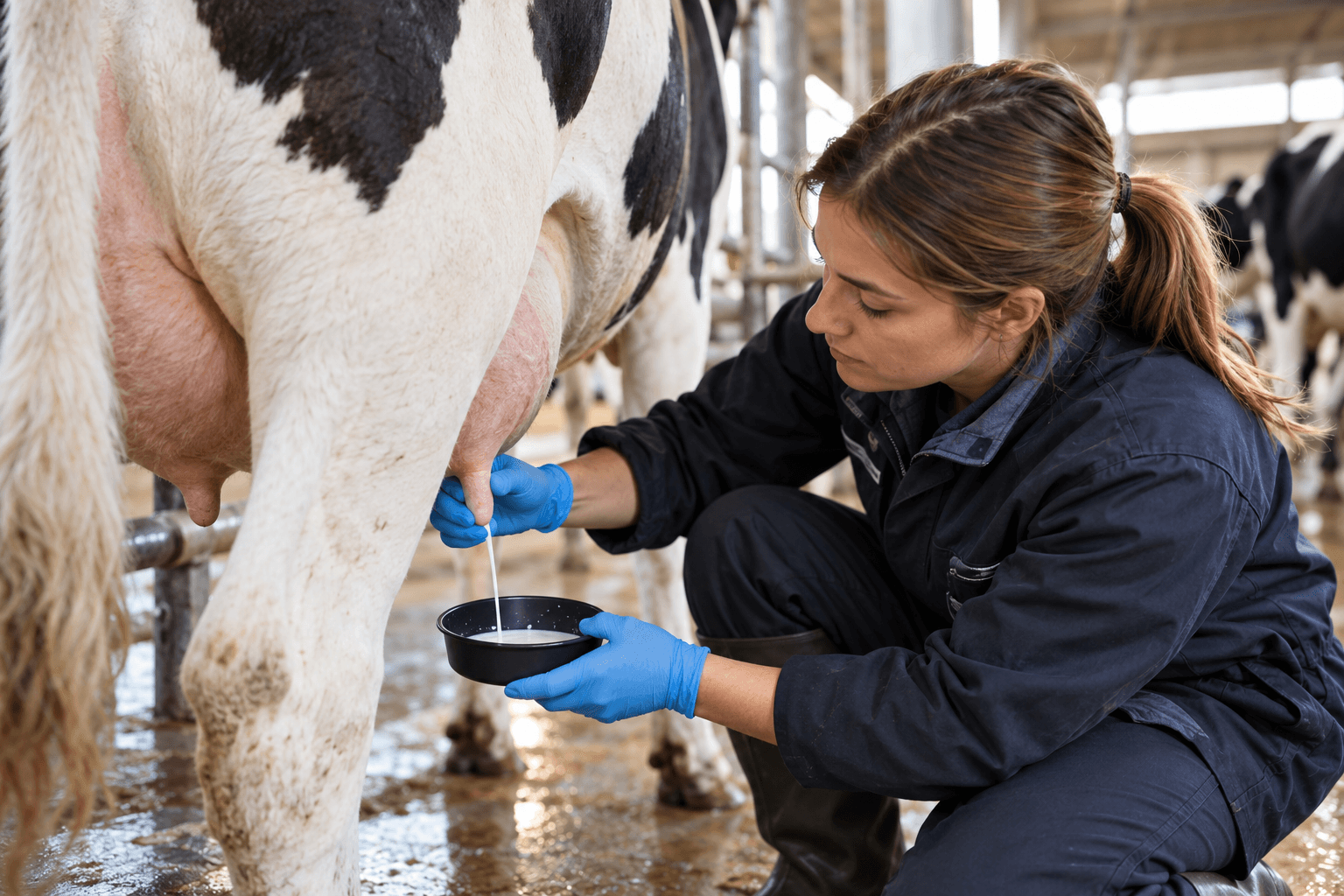
Mastitis is inflammation of the udder, most often caused by bacteria that enter through the teat canal. It is one of the costliest diseases of a dairy herd: the farm loses milk, pays for treatment and gets paid less for the milk it ships. We look at the signs of clinical and subclinical mastitis, its causes, and how it is detected, treated and prevented.
Why mastitis is dangerous for a farm
- Less milk. Yield drops in the affected quarter and often across the whole cow. After severe mastitis, production may not recover until the end of lactation.
- Milk cannot be shipped. During antibiotic treatment and the withdrawal period after it, the cow's milk is discarded.
- Lower price. Mastitis raises the somatic cell count in milk, and dairies pay less for milk with a high count. Under the EAEU technical regulation TR CU 033/2013, raw milk may contain no more than 750,000 somatic cells per ml.
- Culling. Cows with chronic mastitis that does not clear up after treatment have to be culled.
- Spread to other cows. Some pathogens pass from cow to cow during milking.
Clinical and subclinical mastitis
Clinical mastitis is visible to the eye. It is divided into three grades:
- Mild: only the milk has changed: flakes, clots, watery milk.
- Moderate: in addition to changes in the milk, the quarter is swollen, red, firm and painful.
- Severe: the whole cow is sick: fever, refusal to eat, depression. In this case the vet must be called immediately.
Subclinical mastitis cannot be seen: the milk and the udder look normal. But the somatic cell count in the milk rises and yield falls. Subclinical mastitis is 15–40 times more common than clinical mastitis, and it causes most of the farm's losses because it goes unnoticed for a long time.
Causes of mastitis
Mastitis pathogens fall into two groups:
- Contagious: Staphylococcus aureus, Streptococcus agalactiae, mycoplasmas. They live in the udders of infected cows and spread during milking: via the milker's hands, shared towels and teat cups.
- Environmental: E. coli, Klebsiella, Streptococcus uberis. They reach the teats from manure and dirty bedding.
What increases the risk:
- poor milking hygiene: dirty teats, one towel for several cows, working without gloves;
- faulty milking equipment: incorrect vacuum, worn liners, over-milking after milk flow has stopped;
- wet, dirty bedding and cramped stalls;
- the cow lies down right after milking while the teat canal is still open;
- dry-off and the first weeks after calving, when the risk is highest;
- injuries to the teats and udder.
How mastitis is detected
- Forestripping the first streams into a cup with a dark bottom before every milking. This reveals flakes and clots in clinical mastitis.
- A paddle test (the California Mastitis Test, CMT). It shows subclinical mastitis right at the cow, separately for each quarter.
- Somatic cell counting in a laboratory or with an on-farm device. If a cow has more than 200,000 cells per ml of milk, at least one quarter is most likely infected.
- Milk electrical conductivity. Many milking systems and robots measure it for each quarter and warn when it rises.
- Bacterial culture. It shows which pathogen caused the mastitis, and the vet chooses the drug based on it.
Treating mastitis
Treatment is prescribed by a vet. General rules:
- Start as early as possible: the longer the inflammation lasts, the worse the outcome.
- Choose the drug according to the pathogen, not at random. Antibiotics barely work against some pathogens.
- Observe the withdrawal period stated on the drug label. Until it has passed, the cow's milk must not be shipped.
- Record every case: which cow, which quarter, what treatment. This shows which cows get sick repeatedly and which pathogen dominates on the farm.
- A cow with chronic mastitis that returns after treatment infects the others. Her future should be discussed with the vet.
Preventing mastitis
The foundation of prevention is the "five-point plan". It was developed in the UK in the 1960s and has been used by farms around the world ever since:
- Treat and record every clinical case.
- Dip teats with a disinfectant immediately after milking.
- Treat the udder at dry-off according to the vet's protocol.
- Cull cows with chronic mastitis.
- Check and maintain milking equipment regularly.
What else helps:
- clean, dry teats before milking, a separate towel for each cow, gloves for milkers;
- dry, clean bedding;
- fresh feed right after milking so that cows stay standing while the teat canal closes;
- attention to feeding around dry-off and after calving: metabolic problems weaken the udder's defenses.
How cameras and AI help detect mastitis
Milk testing remains the main detection method. Cameras complement it because they see every cow every day. Researchers and developers are trying several approaches:
- Thermal imaging: an inflamed quarter is warmer on the skin surface.
- Regular cameras: udder shape and symmetry, swelling of a single quarter, the cow's behavior during milking, at the feed bunk and in the stalls.
- Milking equipment data: conductivity and yield per quarter.
AI combines these signs and flags the cows that should be tested first.
The FusionAI mastitis module
The FusionAI early mastitis detection module is currently in the pilot stage, and we are recruiting partner farms. The module combines machine vision and an analytics platform to spot signs of udder inflammation earlier and select cows for testing. It does not replace milk tests: the system suggests which cows to check first.
What a pilot farm gets:
- equipment and software on special pilot terms;
- support from the FusionAI team;
- the opportunity to shape the final features of the module.
Other FusionAI modules are already ready to deploy: video-based cow identification without ear tags, early lameness detection, body condition scoring and weighing.
Frequently asked questions
How can I tell if a cow has mastitis?
Clinical mastitis shows in the milk (flakes, clots, watery milk) and in the udder (swelling, redness, firmness, pain). Subclinical mastitis is invisible; it is found with a quick test or by the somatic cell count in the milk.
Why is subclinical mastitis dangerous?
It is 15–40 times more common than clinical mastitis and goes unnoticed for a long time. All this time the cow produces less milk, the somatic cell count rises, and contagious pathogens spread to other cows during milking.
Can milk from a cow with mastitis be shipped?
No. Milk from a cow with clinical mastitis and during treatment must not be shipped. After treatment, only once the withdrawal period stated on the drug label has ended.
Can AI detect mastitis?
A camera cannot see what is happening inside the milk, so milk tests remain the basis. AI helps spot external signs and behavioral changes earlier and suggests which cows to check. FusionAI is currently testing such a module on partner farms.
Want to take part in the mastitis module pilot? Send us a request: we will discuss your farm's needs and the pilot terms.
Related FusionAI solutions
If this topic is relevant for your farm, explore these solutions: